| Home Medical Index | First Posted: Dec 30, 2009 Jan 21, 2020 | |
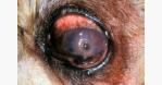
Corneal Abscesses in HorsesSome of the latest research was just released in an abstract Deep lamellar endothelial keratoplasty in 10 horses. Plummer CE, Kallberg ME, Ollivier FJ, Barrie KP, Brooks DE. Department of Small and Large Animal Clinical Sciences, College of Veterinary Medicine, University of Florida, Gainesville, FL 32610-0126, USA. PlummerC@mail. vetmed.ufl.edu OBJECTIVE: To describe and evaluate a surgical technique utilized for the therapy of deep corneal stromal abscesses (DSA) in horses. The DSA is excised and replaced with a partial thickness corneal lamellar allograft. METHODS: A retrospective clinical study describing the indications for the surgical technique utilized and the outcomes of this procedure in 10 eyes of 10 horses. RESULTS: Each affected eye had a discrete DSA within the posterior stroma. An initial partial thickness semicircular corneal incision was made at the limbus, followed by anterior stromal lamellar dissection over the lesion. After excision of the DSA and replacement with a larger diameter split-thickness donor button, the anterior stroma was replaced into its original position and the initial corneal incision was repaired. All of the animals that underwent deep lamellar endothelial keratoplasty (DLEK) procedure healed appropriately and with subjectively less postoperative scarring and complications than previously described surgical approaches to DSA. CONCLUSIONS: This procedure is an effective technique for surgical removal of DSA in horses and, in most cases, results in a visual and cosmetically acceptable globe. The advantages of this technique compared to other surgical approaches to DSA are the peripheral location of the incision, shortened anesthesia times, the resultant minimal scarring and shorter healing times associated with DLEK. Cornea/Superficial Keratitis in the Merck Vet Manual. This link has a tremendous amount of information on horse eye ulcers of all kinds with images. The quoted paragraph, below, is the first paragraph of a comprehensive article. "Superficial keratitis is common in all species and is characterized by corneal vascularization and opacification, which may be due to edema, cellular infiltrates, pigmentation, or fibroplasia. If ulceration is present, pain-manifest by epiphora and blepharospasm-is an outstanding sign. Unilateral keratitis frequently is traumatic in origin. Mechanical factors, such as lid conformational defects and foreign bodies, should always be eliminated as possible causes because improvement will not occur until they are resolved. Ulcerative keratitis may be complicated by secondary invasion by bacteria and, in horses, by saprophytic fungi. Bilateral superficial keratitis may be immune-mediated or associated with a lack of tears, eyelid conformational defects, or infectious agents..." (Follow the above link on Cornea/Superficial Keratitis) Deep Stromal Corneal Ulcer Merck Manual link Deep Stromal Corneal Ulcer
"Deep corneal ulcers, descemetocele, and iris prolapse are seen with some frequency in dogs, cats, and horses. These conditions require immediate surgical support of the weakened cornea as they can threaten or seriously compromise corneal integrity. In dogs, the brachycephalic breeds and dogs with keratoconjunctivitis sicca are most vulnerable. These corneal defects often develop in the center of the cornea and can markedly impair vision. Important diagnostic aids are the Schirmer tear test to measure aqueous tear production and topical fluorescein to examine the corneal ulcer. Corneal culture and cytology can assist in choosing topical and systemic antibiotics. Secondary anterior uveitis with aqueous flare, miosis, ocular hypotony, and hypopyon is common. Corneal ulcer depth must be accurately estimated using magnification, focal illumination, and topical fluorescein. Central corneal ulcers are more vulnerable because they require more time for the healing response and vascularization. Adequate ulcer debridement is essential for successful adherence of a conjunctival graft. The corneal ulceration (stromal, descemetocele, or iris prolapse) is covered with the bulbar conjunctival graft (360 degrees, 180 degrees, bridge, or pedicle) that appears most appropriate. For full-thickness corneal ulcers with iris prolapse, conjunctival grafts are also used, but the postoperative corneal opacity is usually larger and more dense. Postoperative therapy includes topical and systemic broad-spectrum antibiotics, systemic NSAID or corticosteroids, and mydriatics. Treatments are gradually tapered and administered for 4-8 wk. Postoperative complications include variable corneal scar and pigmentation, secondary cataract formation, and rarely, bacterial endophthalmitis." For Further Information: Uveitis |